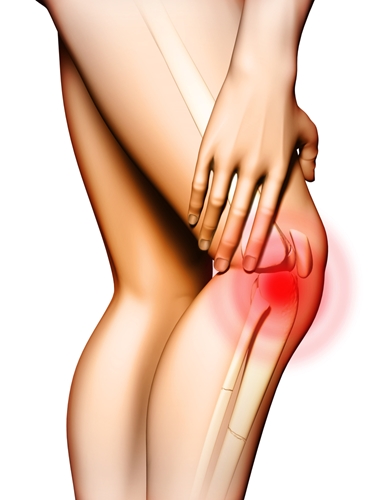
Scientists discover way to reduce effects of traumatic cartilage injuries
While orthopedic devices and state-of-the-art artificial compounds help reduce joint pain, there is little physicians can do once the majority of cartilage breaks down. The lubricating material that covers every bone surface involved in joint manipulation, cartilage does not grow back after traumatic injury or autoimmune responses. It is imperative for patients and physicians alike to be as invested as possible in protecting and maintaining the cartilage that they are born with.
According to a recent study conducted by researchers at the University of Texas at Austin and published in the journal Proceedings of the National Academy of Sciences, the most common way cartilage cells die off has been identified. The scientists hope to leverage their findings into better protective measures for people at risk of developing debilitating arthritis later in life or those who need to preserve what little cartilage their joints may have left.
Protecting cartilage
The University of California, San Francisco explained that the body's cartilage is produced entirely by cells called chondrocytes. Because cartilage is a tough yet flexible material, chondrocytes have a slow time producing it and cannot work fast enough to repair any major rips or tears to cartilage in the elbows, knees or hips. This means that trauma that destroys cartilage may leave irreparable damage.
That is why the Texas researchers looked into the major ways cartilage cells react to traumatic impact events. In a review of how chondrocytes sense pressure-related events, the researchers identified two "Piezo channels" that sense the rapid flow of ions from low- to high-pressure areas. These mechanisms allow chondrocytes to sense both light and hard mechanical forces.
The researchers used synthetic chemicals to block the Piezo channels, and, consequently, chondrocytes' ability to sense pressure and force. The obstruction reduced the prevalence of cell death in controlled laboratory settings, while also making chondrocytes more resistant to overall impact forces.
"The most exciting thing about this study was that it shows that cells in your cartilage, which people don't think of as a typical sensory cell, have multiple sensory systems," Farshid Guilak, M.D., professor of orthopedic surgery at Duke University and contributing author of the study, said in a statement. "These cells are very complex in their ability to sense their mechanical environment."
Guilak explained that while some patients may think that blocking both Piezo channels will make for invincible joints impervious to pain, the pathways allow the body to react appropriately to injury and repair itself if possible. While physicians may one day be able to dampen the effects of traumatic injuries on cell death, completely blocking out the body's natural response could be just as dangerous.
According to the U.S. Centers for Disease Control and Prevention, more than 1 million knee and hip replacements are performed every year. If orthopedic professionals and patients alike are committed to lowering that number, preserving healthy cartilage through further research into Piezo channels may be the most promising option when exercise and orthopedic documentation fails.


